
In cases where a functioning eye becomes damaged or diseased, it is often necessary for the eye to be removed or modified to prepare the eye socket for the fitting of a prosthetic eye. Surgeons that remove or modify eyes are called Ophthalmologists. Surgical specialists within this field are referred to as oculo-plastic surgeons. These doctors will determine if an eye needs to be removed or modified in preparation for a prosthetic eye fitting.
Before The Eye Surgery
Your doctor will instruct you per your diet prior to the surgery. They will give you a period of time when you should not eat before and also advise you to refrain from alcohol or other substances that may interfere with the anesthetics and antibiotics involved during surgery. It is very important to consult with your doctor if you have any questions about diet and preparation before the surgery.
The Eye Surgery
There are generally three types of eye surgery that precede eye prosthesis creation and fitting.
- Enucleation -The removal of the damaged or diseased eyeball.
- Evisceration -The removal of the contents of the eyeball where the outer part (the sclera) remains behind.
- Exenteration -The removal of the entire contents of the orbit, which include removal of the eye muscles.
Enucleation
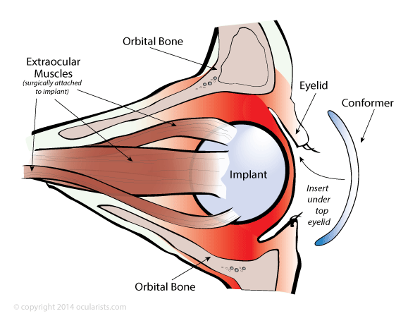
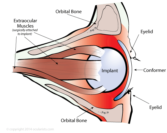
If the eye is removed from the socket (enucleated) then the surgeon places a spherical implant in the eye socket where the eye used to be. The surgeon will also connect the remaining eye muscles to this implant so the implant will move with the companion eye. Once the surgeon has attached the muscles of the eye to the orbital implant, they finalize the surgery by bringing the tissue of the eye (called conjunctiva) across the front of the implant, sewing the conjunctiva together. Since the implant is not as large as the eyeball that was removed, there is space remaining inside the eyelids for the artificial eye to fit. The surgeon attaches the muscles of the eye to the implant so it will have some movement capabilities. The prosthetic eye will be made to fit over this spherical implant and rest underneath the eyelids. Since the eye prosthesis will be custom made to fit over the tissue covering the implant, the eye prosthesis will have some movement as well. An artificial eye is made of a medical grade plastic that is designed to fit within an eye socket after the tissues have healed from surgery. Contrary to popular understanding, eye prostheses are more shell-shaped then ball-shaped since they fit over the sphere implant or existing globe (eye). The surgeon will choose from various types of implants on the market: coral (H.A.), silicone, acrylic or synthetic coral type materials. Each type of material has its own pros and cons. Sometimes dermis-fat grafts are used in place of, or in conjunction with an implant. This subcutaneous fat is harvested from the patient’s own body and is therefore fully bio-compatible. Dermis-fat grafts are primarily used in surgeries of young children since fat may grow and provide stimulus for orbital growth. They are also used in orbital exenteration surgeries. Consult your surgeon if you have questions about the implant option.


Once the surgeon has attached the muscles of the eye to the orbital implant, they finalize the surgery by bringing the tissue of the eye (called conjunctiva) across the front of the implant, sewing the conjunctiva together. Since the implant is not as large as the eyeball that was removed, there is space remaining inside the eyelids for the artificial eye to fit.
Insertion of an eye prosthesis into an enucleated socket

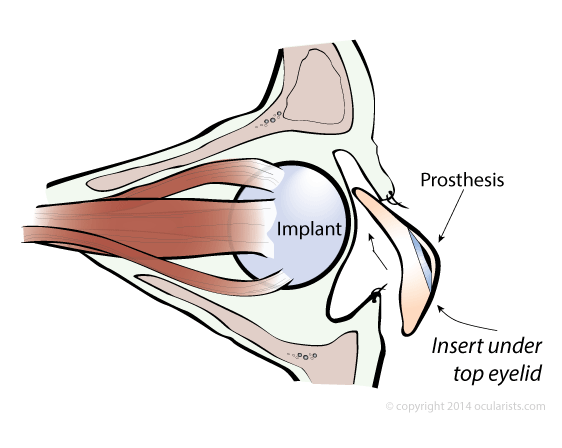

The surgeon attaches the muscles of the eye to the implant so it will have some movement capabilities. The prosthetic eye will be made to fit over this spherical implant and rest underneath the eyelids. Since the eye prosthesis will be custom made to fit over the tissue covering the implant, the eye prosthesis will have some movement as well. An artificial eye is made of a medical grade plastic that is designed to fit within an eye socket after the tissues have healed from surgery. Contrary to popular understanding, eye prostheses are more shell-shaped then ball-shaped since they fit over the sphere implant or existing globe (eye). The surgeon will choose from various types of implants on the market: coral (H.A.), silicone, acrylic or synthetic coral type materials. Each type of material has its own pros and cons. Sometimes dermis-fat grafts are used in place of, or in conjunction with an implant. This subcutaneous fat is harvested from the patient’s own body and is therefore fully bio-compatiable. Dermis-fat grafts are primarily used in surgeries of young children since fat may grow and provide stimulus for orbital growth. They are also used in orbital exenteration surgeries. Consult your surgeon if you have questions about the implant options.
Eye socket after enucleation with implant

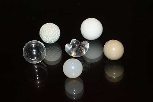
Various types of ocular implants
Evisceration
The removal of the contents of the eyeball where the outer part (the sclera) remains behind is called an evisceration. Sometimes eviscerated eyeballs are filled with an implant. Whether the eviscerated eyeball was filled with an implant or not, the future eye prosthesis will be designed to fit over the remaining eyeball.
Original orbit with eyeball before surgery

Eviscerated eyeball after surgery

Exenteration
In more severe cases, the entire contents of the orbit of the eye are removed, including all the muscles around the eyeball. This is called orbital exenteration surgery. In these cases, an orbital prosthesis will need to be created. An orbital prosthesis is a prosthetic eye with surrounding prosthetic tissue. Like an eye prosthesis, an orbital prosthesis is not a permanent implant and is removable. Microphthalmia Sometimes an eye does not need surgery prior to the fitting of an artificial eye. A microphthalmic eye (small eye) is usually one of these surgical exceptions. Microphthalmia is a congenital condition (at birth) and therefore babies with this condition need to be fitted with a custom fitted prosthesis within the first months of life. If an eye prosthesis or custom conformer (also known as an ocular tissue expander) is not created during infancy, then the child’s face will become asymmetrical as he or she grows. Asymmetry of facial bone and tissue can lead to health problems of the eye socket and sinuses. Therefore, as a child grows, the eye prosthesis is enlarged and replaced to ensure that the bone and tissue grow proportionally.
Entire contents of orbit surgically removed

Cavity with orbital prosthesis

An orbital prosthesis is a prosthetic eye with surrounding prosthetic tissue. Like an eye prosthesis, an orbital prosthesis is not a permanent implant and is removable.
Real Life Examples
|
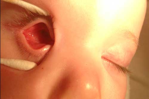
Without Eye Prostheses Patient requiring two eye prostheses. Right eye socket (left in photo) is an enucleated socket with sphere implant. Left eye socket (right in photo) is a phthisical eye (damaged and shrunken eye). |

|
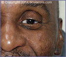
With Eye Prostheses Patient wearing custom eye prostheses in both eye sockets. One in his enucleated socket (left in photo) and one over his phthisical eye (right in photo). |

|
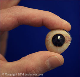
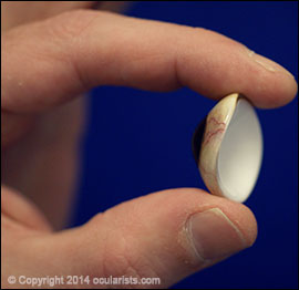
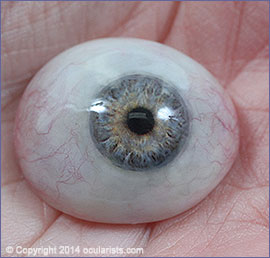
Front View of an Eye Prosthesis Side View of an Eye Prosthesis
|

Example of an orbital prosthesis
Example of an eye prosthesis

Patient with orbital exenteration

Patient wearing orbital prosthesis
Microphthalmia
Sometimes an eye does not need surgery prior to the fitting of an artificial eye. A microphthalmic eye (small eye) is usually one of these surgical exceptions. Microphthalmia is a congenital condition (at birth) and therefore babies with this condition need to be fitted with a custom fitted prosthesis within the first months of life. If an eye prosthesis or custom conformer (also known as an ocular tissue expander) is not created during infancy, then the child’s face will become asymmetrical as he or she grows. Asymmetry of facial bone and tissue can lead to health problems of the eye socket and sinuses. Therefore, as a child grows, the eye prosthesis is enlarged and replaced to ensure that the bone and tissue grow proportionally.
A microphthalmic eye (small eye)
Patient with prosthesis in place


Normal sized eyeball

Microphthalmic Eyeball
Phthisical Eye
Sometimes damaged or diseased eyes (phthisical) do not need to be removed. These cases may still require a prosthesis but one that is very thin. This type of an eye prosthesis is called a scleral-shell eye prosthesis (or haptic lens eye prosthesis).

phthisical eye or damaged eyeball

Phthisical Eye
Phthisical Eye with Prosthesis
Post Surgery
Since the final artificial eye is not ready to be created at the time of surgery, the surgeon places a clear plastic conformer shell in the eye socket. This piece of plastic is called a conformer because it helps conform the socket after surgery as it is healing. It also functions to protect the conjuctival tissue and allow the eyelids to blink without rubbing against the suture line with remaining stitches.
Eye socket with implant after enucleation
Eye socket with conformer in place
The conformer shell sits in the eye socket prior to the creation of a prosthetic eye. It will be worn for six to eight weeks after surgery. Often the doctor will work with the Ocularist to make a temporary artificial eye which functions as a custom conformer.
 DOWNLOAD THIS ARTICLE In PDF Format.